
It is a brain that has won much admiration for the speed and dexterity with which it solves mathematical sums on the quiz show Countdown. Now Carol Vorderman's grey matter has been turned into a work of art and is to go on show.
The telvision celebrity agreed to allow Dr Lythgoe and colleagues at University College London to scan her brain to promote a lecture series run by The Telegraph and the healthcare company Novartis, and funded by the National Endowment for Science, Technology and the Arts, Nesta.
When she read about the experiments on Carol's brain, Miss Palmer contacted Dr Lythgoe because she has been developing a way to create art from scans.

In Carol's case, she etched and marked a series of slices though her brain, taken by the UCL team, on to a stack of glass plates. "I love the end results," said Dr Lythgoe.
"They represent the topography of Carol's mind. I am particularly interested by the ethereal effect of the fragile lines within the solid-but-transparent form, and the spiritual quality of the empty space within which the image seems to float," said Miss Palmer.
"Carol was desperate to know how large her brains were, and was thrilled when I told her they were enormous," she added.
"Amazing," said the presenter, on inspecting the glass versions of her brain for the first time yesterday. She added that she looked forward to having one lit up on her mantlepiece at home.
The result, along with images of Angela Palmer's own brain and that of a convicted murderer, are being shown tonight at at private view in The Fine Art Society in London, before public display tomorrow. One of Carol's brains has already been sold, said Patrick Bourne, managing director.
The inspiration for creating this three dimensional effect came from an exhibit made in 1942 by Britain's only female Nobel prizewinner, Dorothy Hodgkin, said Miss Palmer, who studies at The Ruskin School of Drawing and Fine Art in Oxford.
"She presented the crystal salts of penicillin by drawing contour lines on horizontal perspex sheets, held together by steel bolts."
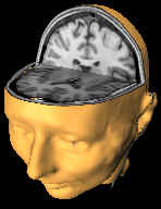
The scans of Carol's brain were made at UCL's Institute of Cognitive Neuroscience by Prof Brian Butterworth and Dr Lythgoe using a method called magnetic resonance imaging.
Image: Eerie glass representations resulted from an unusual collaboration between artist Angela Palmer and scientist Dr Mark Lythgoe, inspired by a competition run by The Telegraph.
Collaborators: Brian Butterworth, Elizabeth Isaacs, Chloe Hutton, Joey Tang, Fulvia Castelli, Hugo Critchley, David Thomas, David Gadian, Roger Ordidge
Structural MRI
At the centre of every hydrogen atom in every water molecule in the human body, there lies a tiny magnet. In everyday life, since these small magnets are weak and relatively very far apart, they are unaware of each other’s presence and point randomly in all directions. However, when the body is placed in the strong magnetic field of an MRI scanner (where the magnetic field is about 30,000 times stronger than the Earth’s natural magnetic field), these tiny magnets line up with the strong field, in the same way a compass needle points to North.
If you push a compass needle away from North, as soon as you let go it will swing back to its original orientation. A similar situation exists for the magnetisation of the human body when it is lying in an MRI scanner. Applying a burst of radio waves while someone is inside the MR scanner causes an electromagnetic force to push the body’s magnetisation out of alignment with the scanner’s magnetic field. Like the compass needle, this is an unnatural state for the body’s magnetisation, and so it quickly returns back to its original position. As it does so, it sends out radio waves, and these are detected and form the signal that makes the final MR image.
Images of the Mind: Functional MRI (fMRI)
Brain activity can be imaged using a technique called functional magnetic resonance imaging (fMRI). fMRI scans of the brain produce images that "light up" when a subject thinks, identifying particular areas of brain activity. When we think, the area of the brain that is working needs more oxygen and the blood supply increases to supply the extra demand. The change in the amount of blood oxygenation causes an increase in the MRI signal intensity where the brain is thinking (coloured area) in the functional MRI image. By acquiring a series of images over time, a picture can be constructed of the activated regions of the brain.
Special thanks: Brian Butterworth, Elizabeth Isaacs, Chloe Hutton, Joey Tang, Fulvia Castelli, Hugo Critchley, David Thomas, David Gadian, Roger Ordidge
Mark Lythgoe and Angela Palmer ©2003